The winners of the 51st annual Nikon Small World Photomicrography Competition have been announced, showcasing some of the most stunning and detailed images of the microscopic world. From insects to plant structures, these photographs reveal the beauty and complexity of tiny subjects we often overlook in our daily lives. The competition, run by Nikon Instruments, has been celebrating excellence in microscopy and digital imaging for over five decades, inspiring scientists and artists alike.
This year’s top prize went to China’s Zhang You for his incredible image of a rice weevil perched on a grain of rice, wings fully extended. Using a combination of photography skills, careful lighting, and focus stacking, You captured a moment that blends scientific detail with artistic flair. His work reminds us that even the smallest creatures can be fascinating and beautiful when seen up close.
More info: nikonsmallworld.com | Instagram | Facebook | x.com
#1 Image Of Distinction: Ye Fei Zhang, Jiang Yin, Jiang Su, China
“Butterfly (Artopoetes pryeri) eggs.”
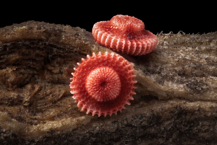
Image credits: © Ye fei Zhang | Nikon Small World
Zhang You didn’t just win first place; he also earned 15th place with a second image showing a Geometer moth laying eggs. A member of the Entomological Society of China, You has spent years studying insects and teaching others about them. He says the key to a great microscopic photo is a mix of science and art, from understanding the subject’s behavior to mastering lighting. “It pays to dive deep into entomology: understanding insects’ behaviors and mastering lighting,” You said. “A standout work blends artistry with scientific rigor, capturing the very essence, energy, and spirit of these creatures.”
#2 1st Place: Zhang You, Kunming, Yunnan, China
“Rice weevil (Sitophilus oryzae) on a grain of rice.”
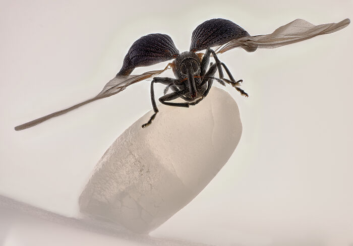
Image credits: © Zhang You | Nikon Small World
#3 Honorable Mention: Michael Robert Peres, Rochester Institute Of Technology, School Of Photographic Arts And Sciences, Rochester, New York, USA
“Melting snowflake.”
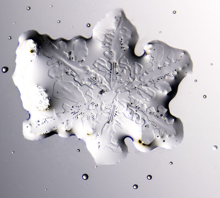
Image credits: © Michael Robert Peres | Nikon Small World
“Zhang You’s work demonstrates the remarkable power of microscopy to reveal new perspectives on the world around us,” said Eric Flem, Senior Manager, Communications and CRM at Nikon Instruments. “What makes this year even more extraordinary is that it was his very first time entering the competition, and he not only captured first place, but also placed another image in the top 20. His achievement highlights the spirit of Nikon Small World: inspiring wonder, making scientific understanding accessible to all, and celebrating the artistry of the microscopic realm.”
#4 Image Of Distinction: Daniel Evrard, Aywaille, Liege, Belgium
“Androconial (pheromone producing) area of a butterfly (Colias) wing.”
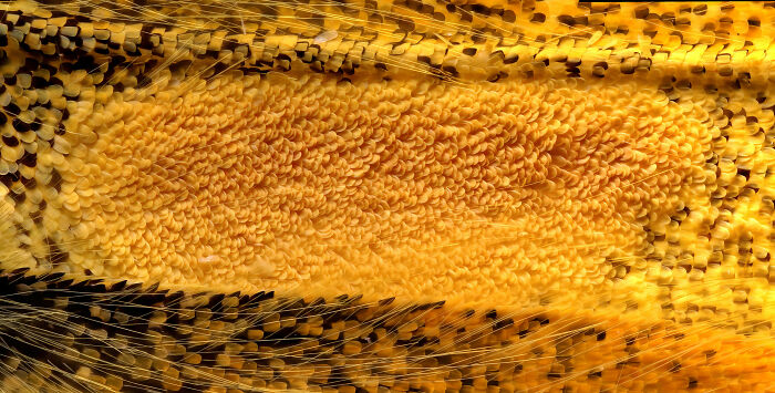
Image credits: © Daniel Evrard | Nikon Small World
#5 Image Of Distinction: Dr. Francisco Lázaro-Diéguez, Albert Einstein College Of Medicine, Bronx, New York, USA
“Dedifferentiated liver cell.”
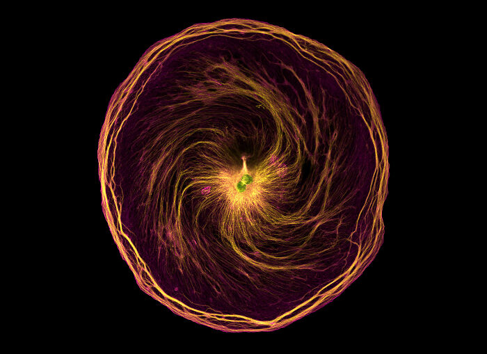
Image credits: © Dr. Francisco Lázaro-Diéguez | Nikon Small World
The second-place winner, Dr. Jan Rosenboom from Germany, captured stunning spheres of Volvox algae in a drop of water, while third place went to John-Oliver Dum, also from Germany, for an intricate photo of pollen caught in a garden spider’s web. Both images reveal patterns and details that most people would never notice in everyday life.
#6 Image Of Distinction: Joe Mckellar, Cnrs, The Institue Of Molecular Genetics Of Montpellier (Igmm), Montpellier, Hérault, France
“Giant human hepatic cancer cell surrounded by smaller cells.”

Image credits: © Joe Mckellar | Nikon Small World
#7 Image Of Distinction: Dr. Michael Weber, Berlin Institute Of Health At Charité, Department Of Human Genetics, Berlin, Germany
“Blood vessels in the limb of an embryonic mouse.”

Image credits: © Dr. Michael Weber | Nikon Small World
Many of the top images use a technique called “image stacking,” where multiple photos are combined to create one sharp, detailed picture. Others rely on confocal or fluorescence microscopy to highlight structures inside cells or tiny organisms, letting viewers see hidden shapes and colors that would otherwise be invisible.
#8 Image Of Distinction: Robert Schmittling, Hillsborough, North Carolina, USA
“Eye of potato (stomate).”

Image credits: © Robert Schmittling | Nikon Small World
#9 Honorable Mention: Jiri Cerny, Institute Of Molecular Genetics Of The Czech Academy Of Sciences, Light Microscopy Core Facility, Prague, Czech Republic
“Jumping spider.”
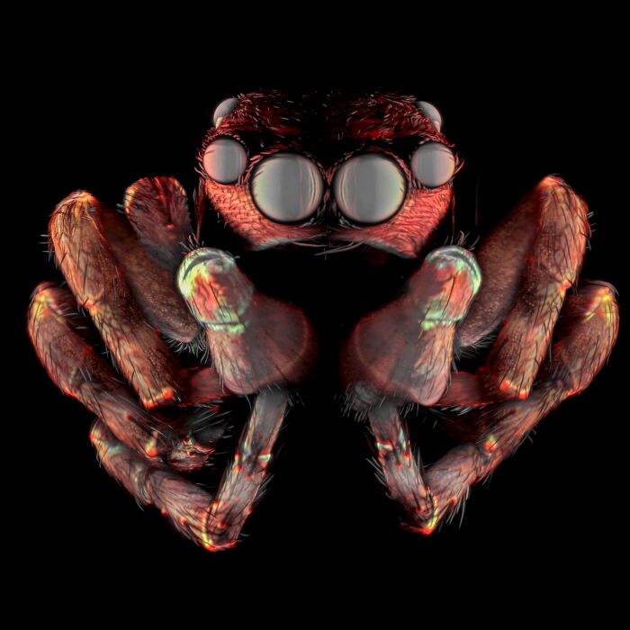
Image credits: © Jiri Cerny | Nikon Small World
In total, the competition recognized 71 images from thousands of entries worldwide. From crystallized soy sauce to mouse neurons, the photos show the incredible variety of life under the microscope—and how curiosity and patience can turn even the tiniest subjects into breathtaking works of art.
#10 Honorable Mention: Gregory B. Murray, Pritchard, British Columbia, Canada
“Frost on a wooden railing.”
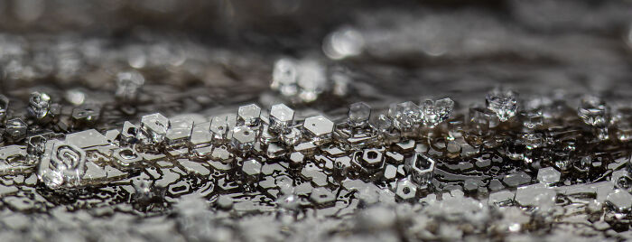
Image credits: © Gregory B. Murray | Nikon Small World
#11 Honorable Mention: Dr. Bruno Cisterna And Dr. Eric Vitriol, Medical College Of Georgia At Augusta University, Department Of Neuroscience & Regenerative Medicine, Augusta, Georgia, USA
“Human neurons reprogrammed from skin cells.”
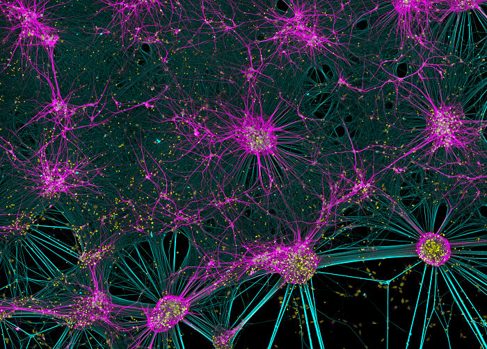
Image credits: © Dr. Bruno Cisterna and Dr. Eric Vitriol | Nikon Small World
#12 Honorable Mention: Mishal Abdulaziz Alryhan, Fiap, Al-Ahsa, Saudi Arabia
“Crystallized soy sauce fusion with alum.”
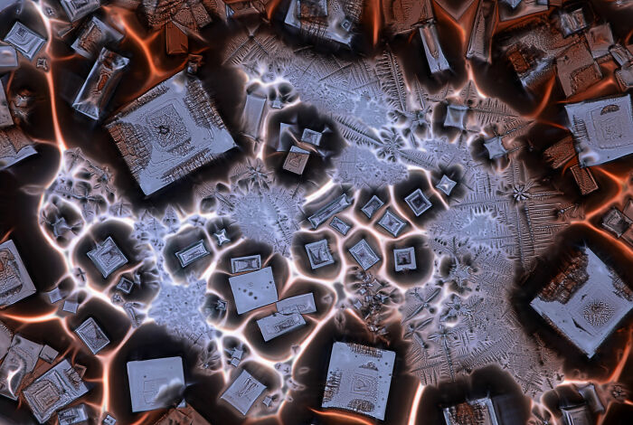
Image credits: © Mishal Abdulaziz Alryhan | Nikon Small World
#13 Image Of Distinction: Jianguo Mao, Shanghai, Shanghai, China
“Pregnant water flea (Daphnia).”
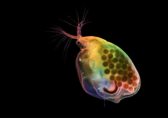
Image credits: © Jianguo Mao | Nikon Small World
#14 Image Of Distinction: Karl Deckart, Eckental, Bavaria, Germany
“Recrystallization of phenyl imidazol.”

Image credits: © Karl Deckart | Nikon Small World
#15 Image Of Distinction: Dr. Noah Bressman, Salisbury University, Department Of Biology, Salisbury, Maryland, USA
“Histologically-stained harvestfish/star butterfish (Peprilus paru).”
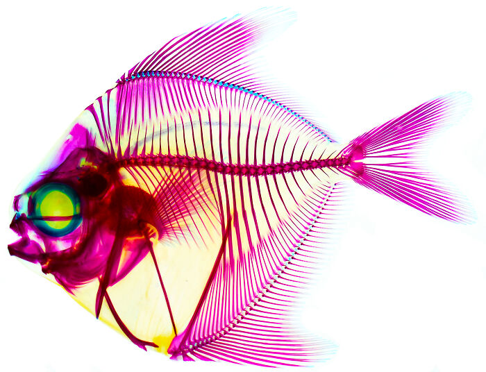
Image credits: © Dr. Noah Bressman | Nikon Small World
#16 Image Of Distinction: Walter Ferrari, Walter Ferrari Macro, Rio Cuarto, Cordoba, Argentina
“True bug (Hemipteran) eggs on a leaf.”
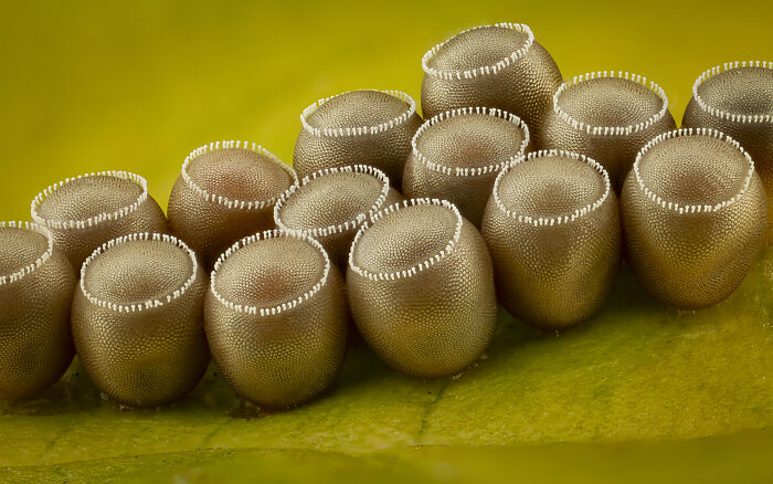
Image credits: © Walter Ferrari | Nikon Small World
#17 3rd Place: John-Oliver Dum, Medienbunker Produktion, Bendorf, Rheinland Pfalz, Germany
“Pollen in a garden spider web.”
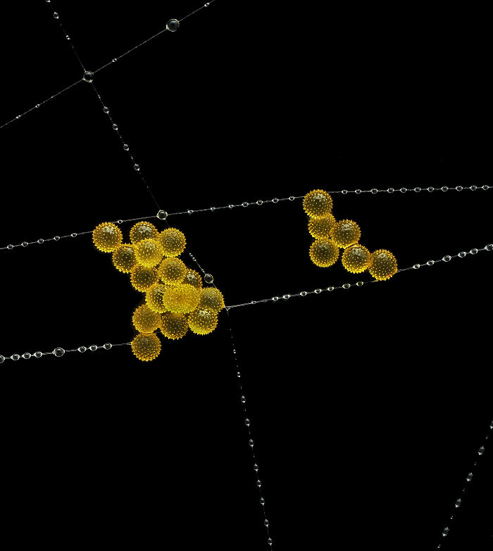
Image credits: © John-Oliver Dum | Nikon Small World
#18 9th Place: Wim Van Egmond, Micropolitan Museum, Berkel En Rodenrijs, Zuid Holland, Netherlands
“A fungus (Talaromyces purpureogenus) known for its red, diffused pigment.”
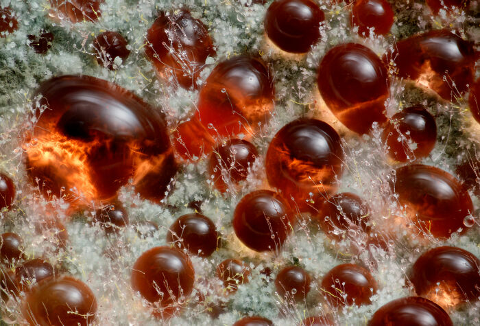
Image credits: © Wim van Egmond | Nikon Small World
#19 11th Place: Marek Miś, Marek Miś Photography, Suwalki, Podlaskie, Poland
“Sunflower trichomes (hair-like plant outgrowths).”
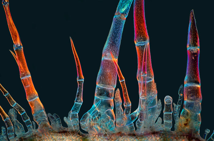
Image credits: © Marek Miś | Nikon Small World
#20 Honorable Mention: Rebecca Lee, Yale University, Department Of Genetrics, New Haven, Connecticut, USA
“Villi in the mouse small intestine.”
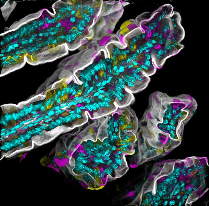
Image credits: © Rebecca Lee | Nikon Small World
#21 Honorable Mention: Michael Parra Puentes, Bogotá, Cundinamarca, Colombia
“Thoracic and cephalic horn of a male beetle (Golofa porteri).”
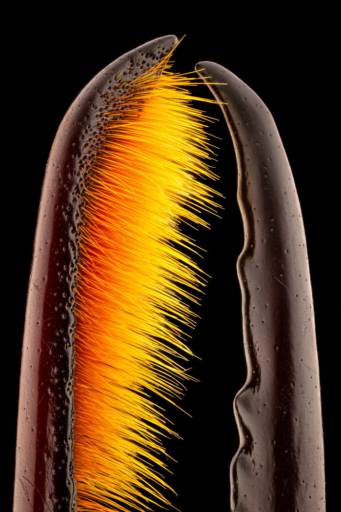
Image credits: © Michael Parra Puentes | Nikon Small World
#22 Honorable Mention: Kendall O. Myers And Dr. Matthew S. Lehnert, Kent State University At Stark, Department Of Biological Sciences, North Canton, Ohio, USA
“Hook-like crochets on the larva of an Io (Automeris io) moth.”
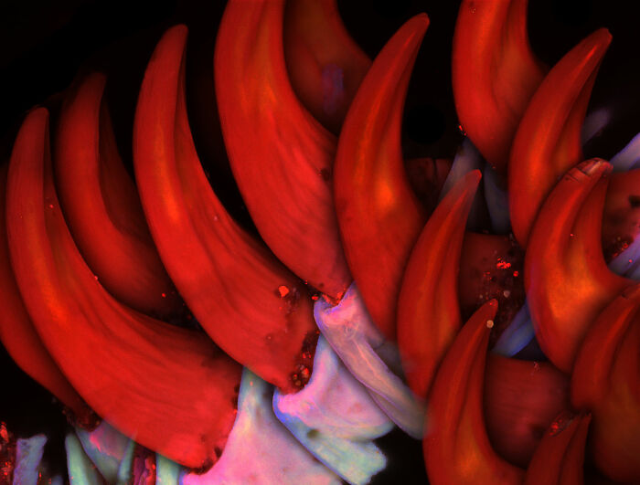
Image credits: © Kendall O. Myers and Dr. Matthew S. Lehnert | Nikon Small World
#23 Image Of Distinction: Dr. Amy C. Engevik, Medical University Of South Carolina, Department Of Regenerative Medicine & Cell Biology, Charleston, South Carolina, USA
“Mouse small intestine.”
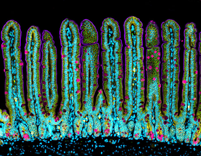
Image credits: © Dr. Amy C. Engevik | Nikon Small World
#24 Image Of Distinction: Dr. Arthur Chien And Dr. Ann Na Cho, Macquarie University, Microscopy Facility, Maff, Macquarie University, New South Wales, Australia
“3D brain organoids in a custom organ-on-a-chip device.”
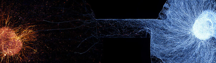
Image credits: © Dr. Arthur Chien and Dr. Ann Na Cho | Nikon Small World
#25 Image Of Distinction: Bernard Allard, Club Français De Microscopie, Sucy-En-Bry, France
“Parasitic fly (Crataerina hirundinis).”
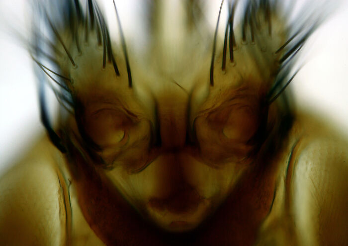
Image credits: © Bernard Allard | Nikon Small World
#26 Image Of Distinction: Bilal Akhtar, Institute Of Molecular Biology, Neuroscience-Rna Biology, Mainz, Rheinland-Pfalz, Germany
“14-day-old mouse neuronal co-culture with astrocytes.”
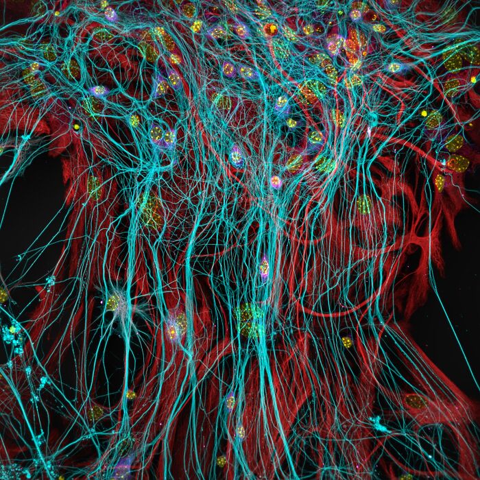
Image credits: © Bilal Akhtar | Nikon Small World
#27 Image Of Distinction: Dr. David Maitland, Art Of Science, St. Andrews, Fife, United Kingdom
“Vascular bundles in a bamboo leaf (Phyllostachys sp.).”
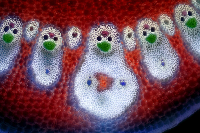
Image credits: © Dr. David Maitland | Nikon Small World
#28 Image Of Distinction: Frederic Labaune, Education Nationale, Auxonne, Burgundy, France
“Slime mold (Arcyria denudata).”
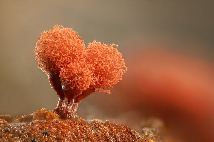
Image credits: © Frederic Labaune | Nikon Small World
#29 Image Of Distinction: Hannah Somers, Mdi Biological Laboratory, Light Microscopy Facility, Bar Harbor, Maine, USA
“An adult zebrafish showing blood vessels in the brain.”
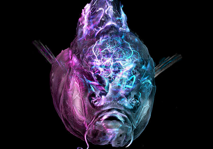
Image credits: © Hannah Somers | Nikon Small World
#30 Image Of Distinction: Heiti Paves, Tallinn, Harju, Estonia
“Mouse embryo, sagittal section.”
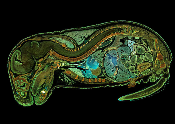
Image credits: © Heiti Paves | Nikon Small World
#31 Image Of Distinction: Jean-Marc Babalian, Nantes, France
“3/4 view of an old Pentium 90 processor.”
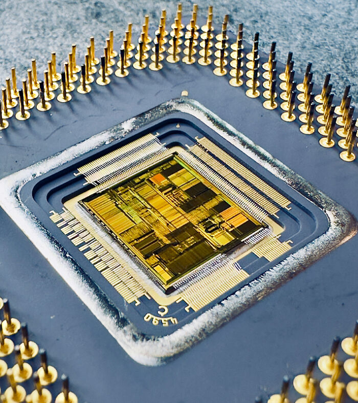
Image credits: © Jean-marc Babalian | Nikon Small World
#32 Image Of Distinction: Dr. Julien Resseguier, University Of Oslo, Department Of Biosciences / Fyscell, Oslo, Viken, Norway
“Immune cells (magenta) protecting the different tissue compartments of the zebrafish intestines.”
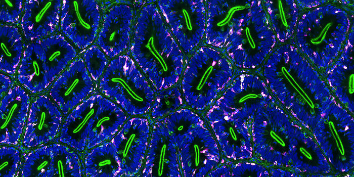
Image credits: © Dr. Julien Resseguier | Nikon Small World
#33 Image Of Distinction: Lauren Johnson, Powered Research, In Vitro Services, Durham, North Carolina, USA
“Mouse retina showing vasculature (red), nerve bundles (green) and macrophages (magenta).”
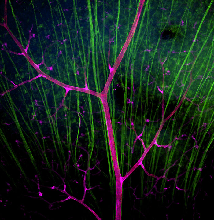
Image credits: © Lauren Johnson | Nikon Small World
#34 Image Of Distinction: Dr. Laurent Formery, University Of California Berkeley, Department Of Molecular And Cell Biology, Pacific Grove, California, USA
“Skeleton of a juvenile sea cucumber.”
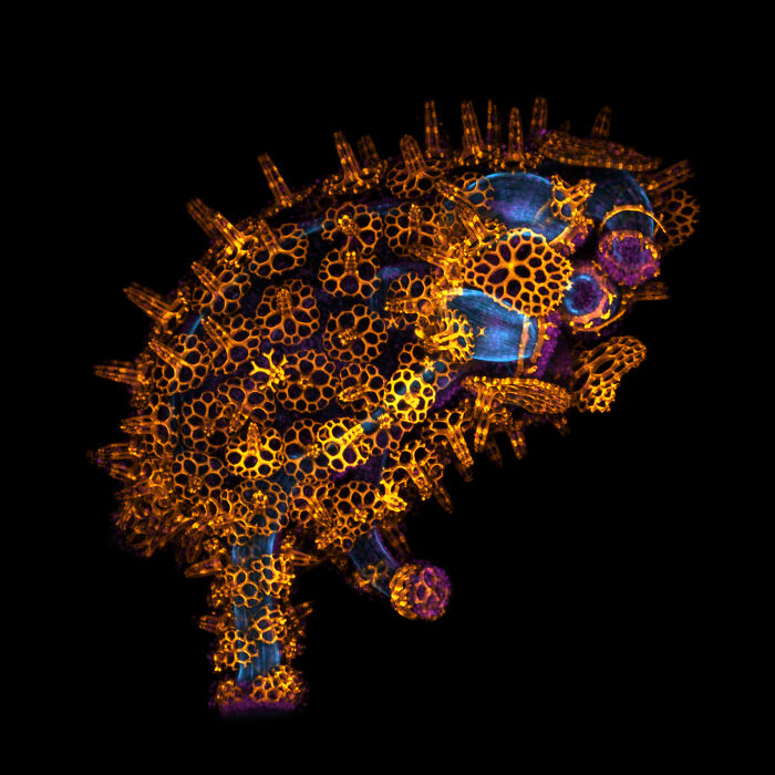
Image credits: © Dr. Laurent Formery | Nikon Small World
#35 Image Of Distinction: Marek Miś, Marek Miś Photography, Suwalki, Podlaskie, Poland
“Air bubbles in melted polyvinyl alcohol.”

Image credits: © Marek Miś | Nikon Small World
#36 Image Of Distinction: Marek Miś, Marek Miś Photography, Suwalki, Podlaskie, Poland
“Crystallized soy sauce.”
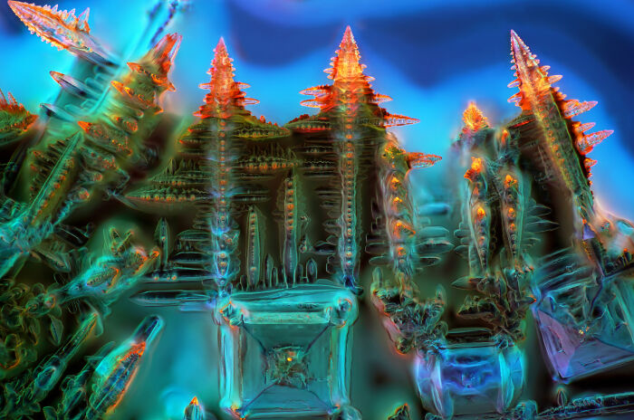
Image credits: © Marek Miś | Nikon Small World
#37 Image Of Distinction: Dr. Mette Handberg-Thorsager, Alexandre Alié And Lisa Maria Ulbrich, Georg-August-University Göttingen, Department Of Multiscale Biology, Göttingen, Niedersachsen, Germany
“Oozoid of a sea squirt (Thalia democratica).”
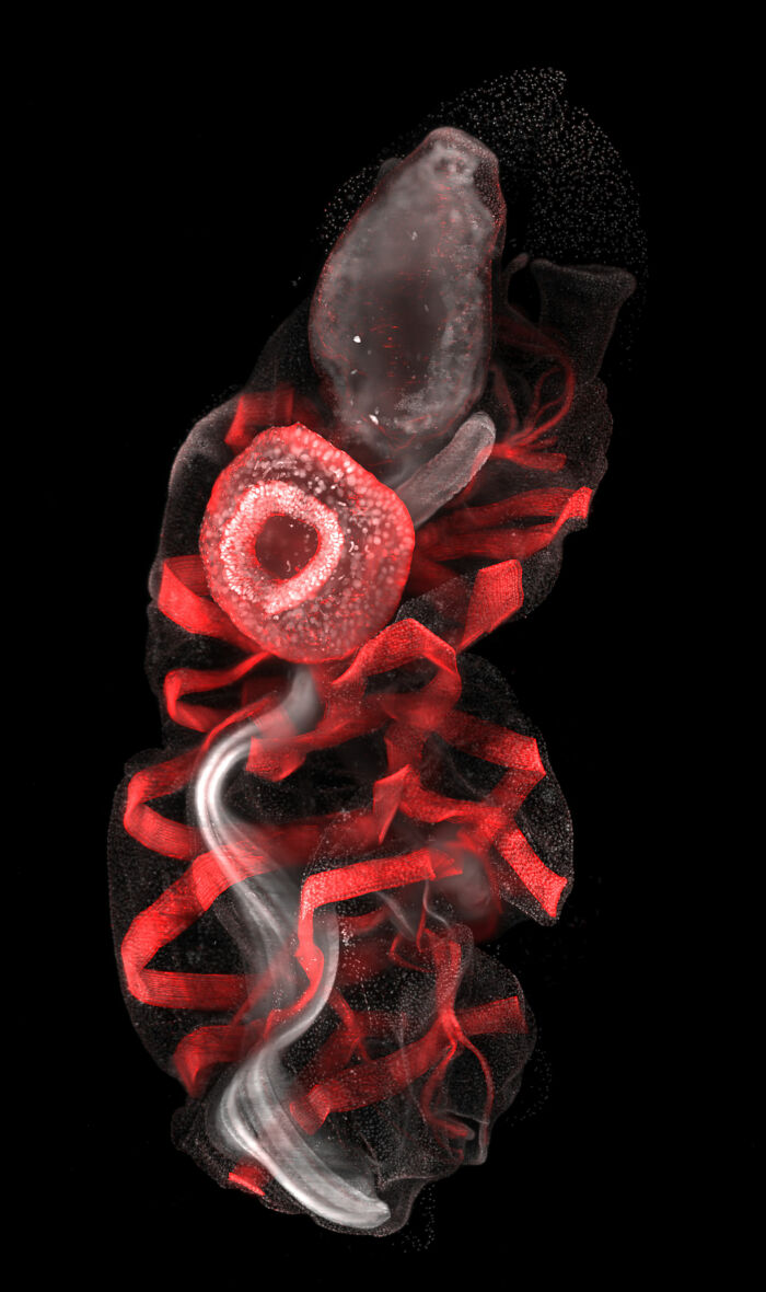
Image credits: © Dr. Mette Handberg-Thorsager, Alexandre Alié and Lisa Maria Ulbrich | Nikon Small World
#38 Image Of Distinction: Özgür Kerem Bulur, Istanbul, Turkey
“Spotted eye hoverfly.”
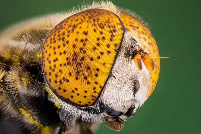
Image credits: © Özgür kerem Bulur | Nikon Small World
#39 Image Of Distinction: Dr. Rory L. Cooper And Professor Michel Milinkovitch, University Of Geneva, Department Of Genetics And Evolution, Geneva, Switzerland
“Wing of the chicken embryo after 11 days of development.”
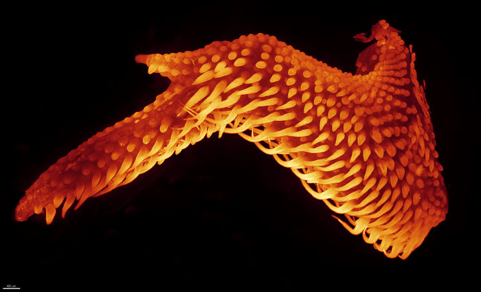
Image credits: © Dr. Rory L. Cooper and Professor Michel Milinkovitch | Nikon Small World
#40 Image Of Distinction: Solvin Zankl, Kiel, Schleswig-Holstein , Germany
“A floating sea slug (Glaucus atlanticus, also known as the blue sea dragon).”
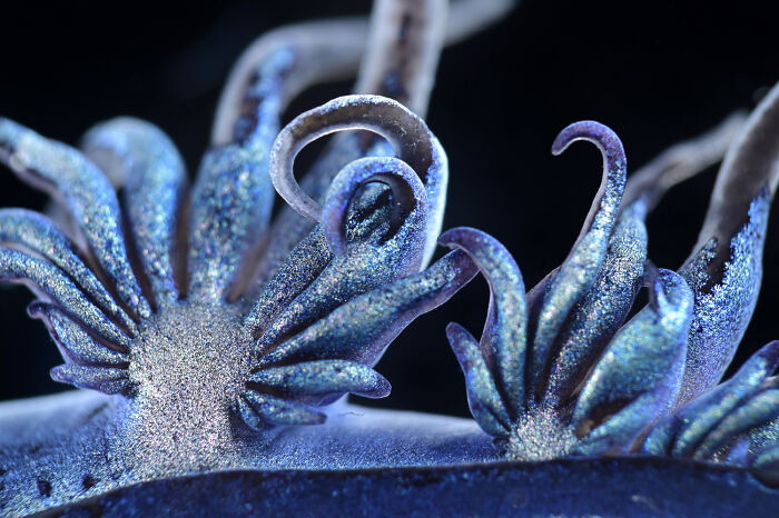
Image credits: © Solvin Zankl | Nikon Small World
#41 Image Of Distinction: Dr. Stephen De Lisle, Karlstad University, Department Of Environmental And Life Sciences, Karlstad, Värmland, Sweden
“Lily flower pollen (autofluorescence).”
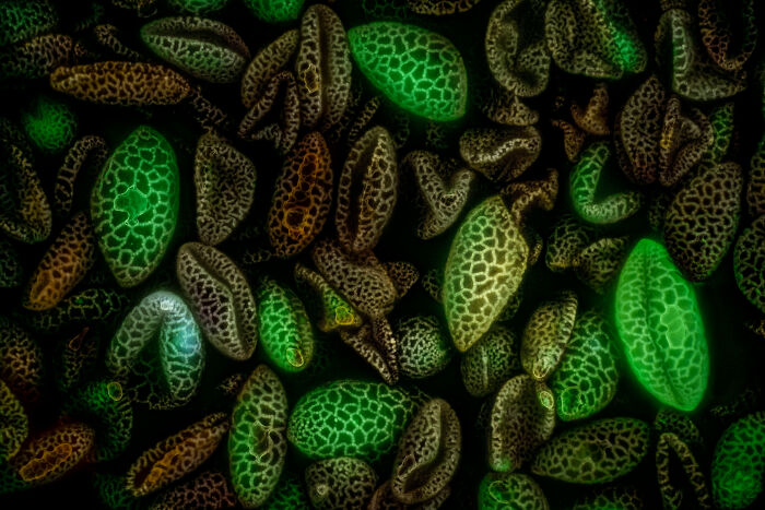
Image credits: © Dr. Stephen de Lisle | Nikon Small World
#42 Image Of Distinction: Dr. Stephen De Lisle, Karlstad University, Department Of Environmental And Life Sciences, Karlstad, Värmland, Sweden
“Planktonic microalgae (Dinobryon).”
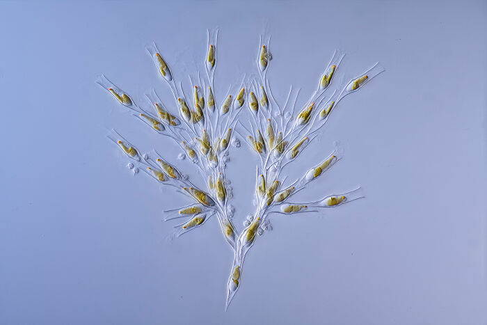
Image credits: © Dr. Stephen de Lisle | Nikon Small World
#43 2nd Place: Dr. Jan Rosenboom, Rostock, Mecklenburg Vorpommern, Germany
“Colonial algae (Volvox) spheres in a drop of water.”
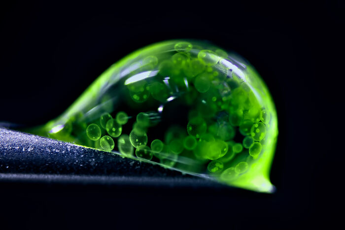
Image credits: © Dr. Jan Rosenboom | Nikon Small World
#44 4th Place: Dr. James Hayes, Vanderbilt University, Department Of Cell And Developmental Biology, Nashville, Tennessee, USA
“Heart muscle cells with chromosomes condensed following cell division.”

Image credits: © Dr. James Hayes | Nikon Small World
#45 5th Place: Dr. Igor Siwanowicz, Howard Hughes Medical Institute (Hhmi), Janelia Research Campus, Ashburn, Virginia, USA
“Spores (blue/purple structures) of a small tropical fern (Ceratopteris richardii).”
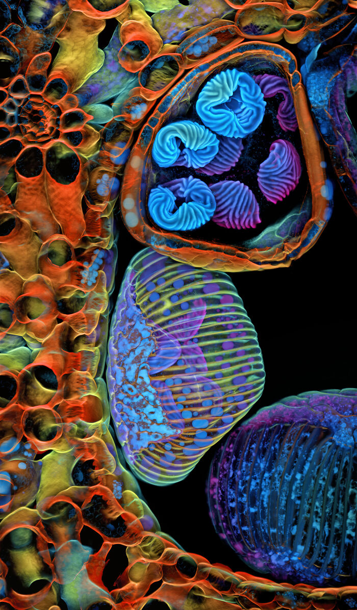
Image credits: © Dr. Igor Siwanowicz | Nikon Small World
#46 6th Place: Dr. Francisco Lázaro-Diéguez, Albert Einstein College Of Medicine, Bronx, New York, USA
“Rat liver cells.”
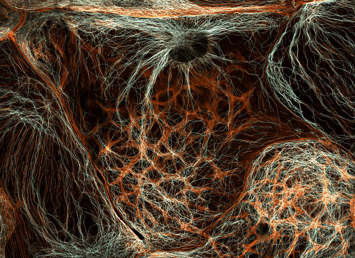
Image credits: © Dr. Francisco Lázaro-Diéguez | Nikon Small World
#47 8th Place: Dr. Igor Siwanowicz, Howard Hughes Medical Institute (Hhmi), Janelia Research Campus, Ashburn, Virginia, USA
“Mallow pollen germinating on stigma while being parasitized by a filamentous fungus.”
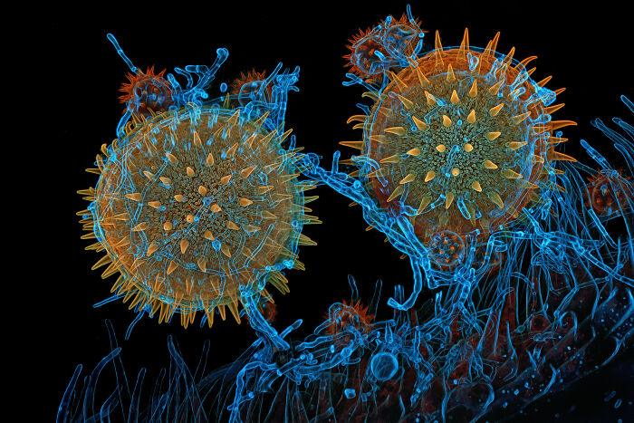
Image credits: © Dr. Igor Siwanowicz | Nikon Small World
#48 10th Place: Dr. Dylan Burnette And Dr. James Hayes, Vanderbilt University School Of Medicine, Department Of Cell And Developmental Biology, Nashville, Tennessee, USA
“Heart muscle cells (iPSC-derived) showing condensed chromosomes in metaphase.”

Image credits: © Dr. Dylan Burnette and Dr. James Hayes | Nikon Small World
#49 13th Place: Henri Koskinen, Helsinki University, Helsinki, Uudenmaan Lääni, Finland
“Slime mold (Arcyria major) releasing spores.”
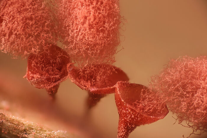
Image credits: © Henri Koskinen | Nikon Small World
#50 14th Place: Manfred Heising, Lwl Museum Of Natural History Münster, Münster, Northrhine-Westphalia, Germany
“Quartz with biotic goethite filaments.”
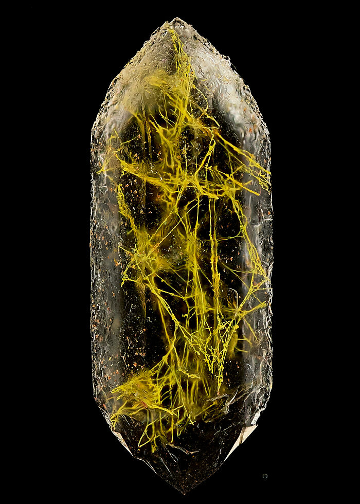
Image credits: © Manfred Heising | Nikon Small World
#51 15th Place: Zhang You, Kunming, Yunnan, China
“Geometer moth (Geometridae) laying eggs.”
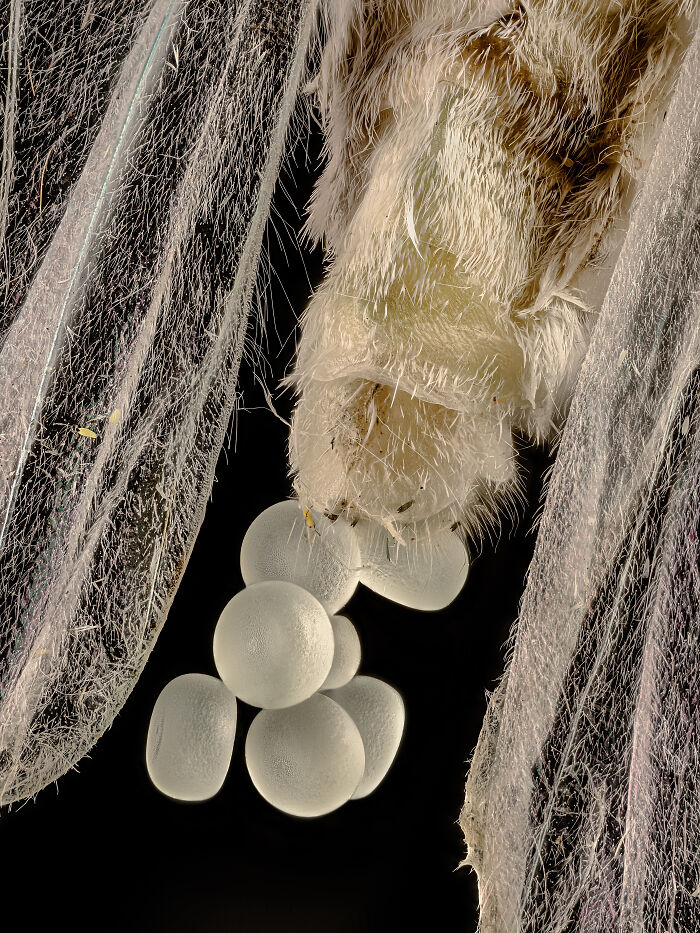
Image credits: © Zhang You | Nikon Small World
#52 16th Place: Rogelio Moreno, Panama, Panama
“Spore sacs (sporangia) of a fern.”
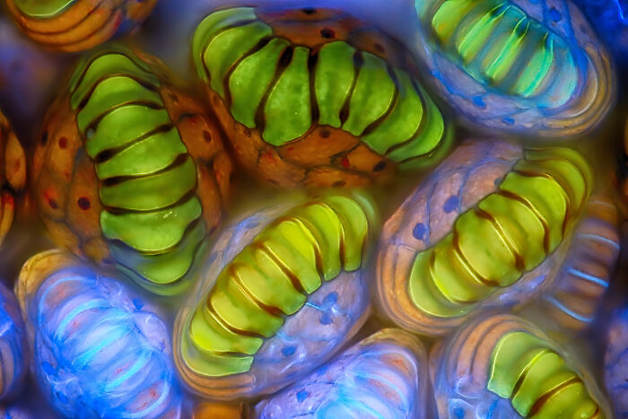
Image credits: © Rogelio Moreno | Nikon Small World
#53 19th Place: Eduardo Carrasco, Cuenca, Azuay, Ecuador
“Parasitic fungus (Cordycipitaceae) on a fly (Calliphoridae).”
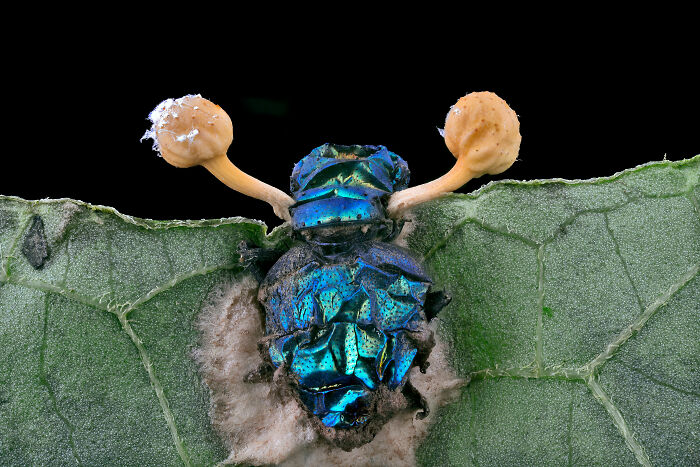
Image credits: © Eduardo Carrasco | Nikon Small World
#54 20th Place: Zachary Sanchez, Vanderbilt University, Department Of Cell And Developmental Biology, Nashville, Tennessee, USA
“Marine copepod .”
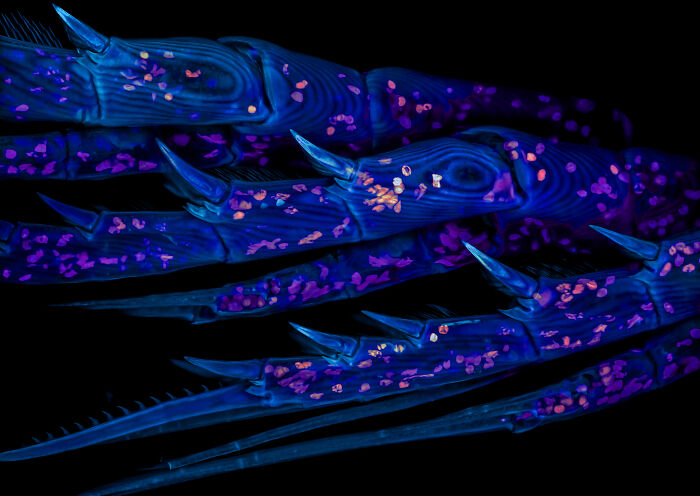
Image credits: © Zachary Sanchez | Nikon Small World
#55 Honorable Mention: Dr. Frédéric Fercoq And Jean-Gabriel Rothan, Muséum National D’histoire Naturelle, Paris, France
“Larvae of a filarial parasite (nematode).”
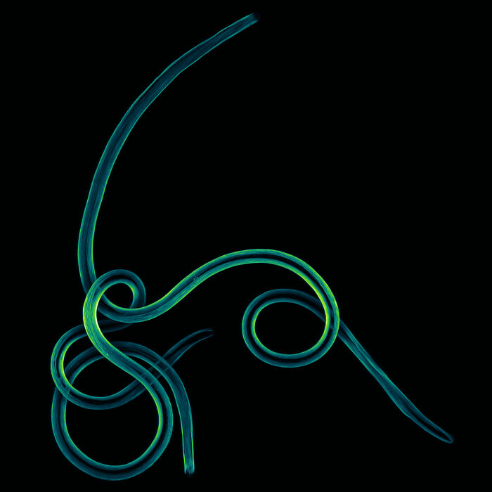
Image credits: © Dr. Frédéric Fercoq and Jean-Gabriel Rothan | Nikon Small World
#56 Honorable Mention: Dr. Zisong Ma, University Of Science And Technology Of China, Hefei, Anhui, China
“Corydalis pallida seed (light yellow) and elaiosome droplet (semitransparent).”
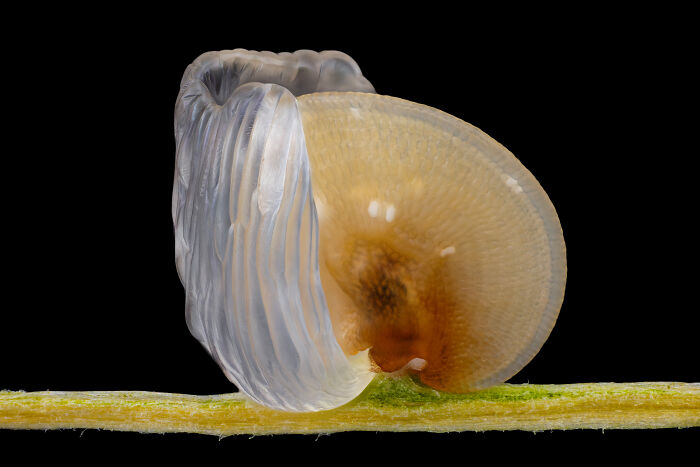
Image credits: © Dr. Zisong Ma | Nikon Small World
#57 Image Of Distinction: Charles Krebs, Charles Krebs Photography, Issaquah, Washington, USA
“Barnacle cirri exoskeleton auto-fluorescing. Diatoms with chlorophyll are shown in bright red.”
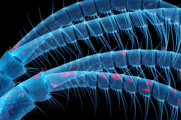
Image credits: © Charles Krebs | Nikon Small World
#58 Image Of Distinction: Daniel Han, Diatoms Australia (Macro Cosmos Imaging), Sydney, New South Wales, Australia
“Diatoms (Arachnoidiscus sp.) on coralline algae.”
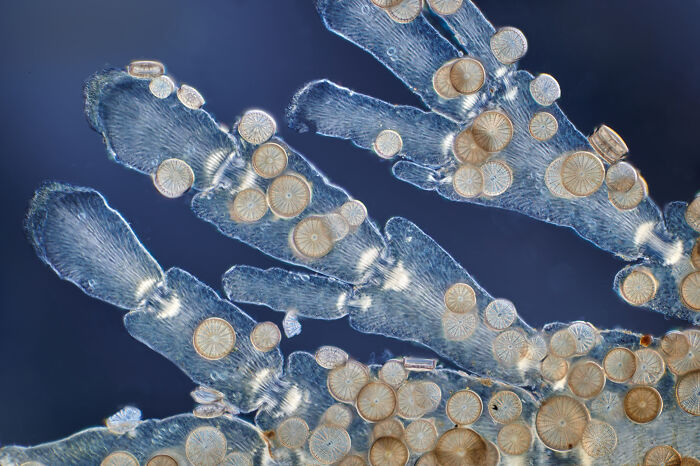
Image credits: © Daniel Han | Nikon Small World
#59 Image Of Distinction: Doong Yien, Xmato Works, Beijing, China
“Crystallization of a mixed solution of alanine and glutamine under polarized light.”
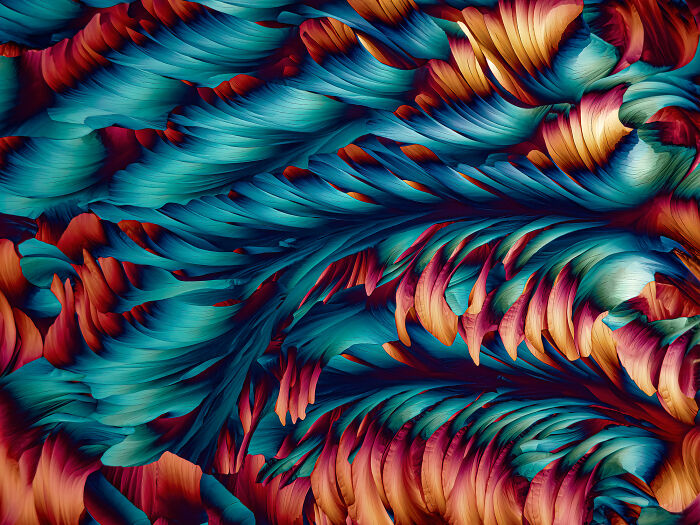
Image credits: © Doong Yien | Nikon Small World
#60 Image Of Distinction: Frantisek Bednar, Svosov, Zilinsky, Slovak Republic
“Filamentous green alga (Spirogyra sp.) showing conjugating tubes and fused cells (zygotes).”
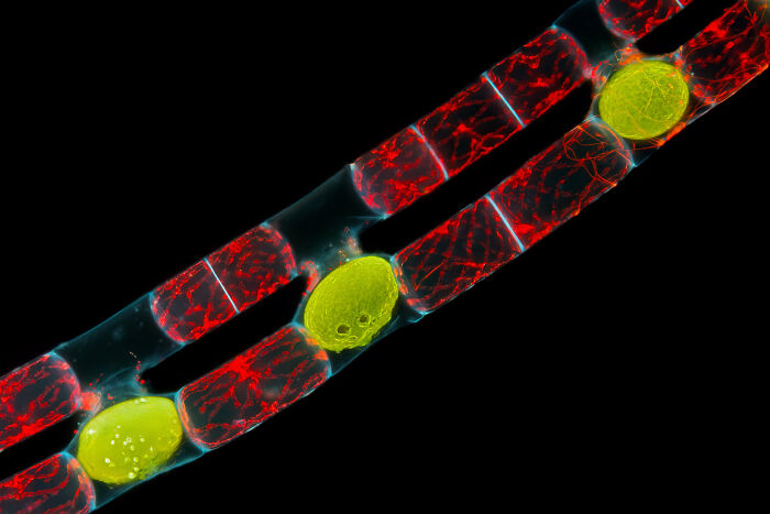
Image credits: © Frantisek Bednar | Nikon Small World
#61 Image Of Distinction: Dr. Gonzalo Quiroga Artigas, Crbm-Cnrs, Montpellier, Herault, France
“Tardigrade.”
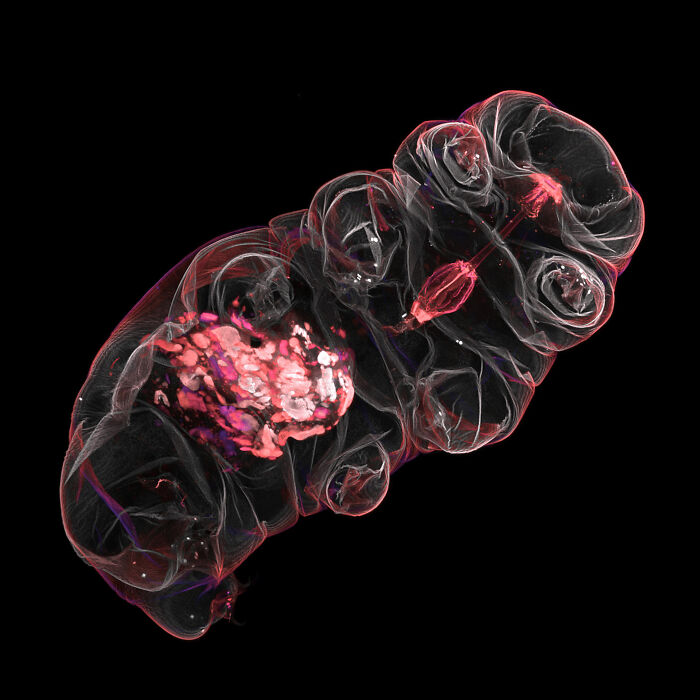
Image credits: © Dr. Gonzalo Quiroga Artigas | Nikon Small World
#62 Image Of Distinction: Igor Rudkovsky
“Slime mold (Cribraria purpurea).”
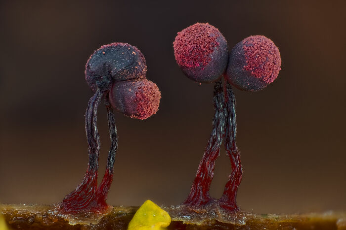
Image credits: © Igor Rudkovsky | Nikon Small World
#63 Image Of Distinction: Jonathan Muyal, Paris, France
“Iridescent rutile (mineral) needles in a Burmese ruby.”

Image credits: © Jonathan Muyal | Nikon Small World
#64 Image Of Distinction: Shambhavi Dwivedi And Dr. Friedemann Kiefer, University Of Münster, Faculty Of Biology, Muenster, North-Rhine Westphalia, Germany
“Mouse lymphatic network (red) flanking blood vessels (white).”
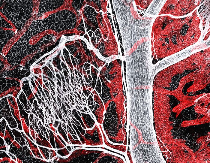
Image credits: © Shambhavi Dwivedi and Dr. Friedemann Kiefer | Nikon Small World
#65 Image Of Distinction: Syed Ashraf, Dr. Divya Sridharan And Ms. Salvia Zafar, The Ohio State University, Department Of Emergency Medicine, Columbus, Ohio, USA
“Human iPSC-derived cardiac organoid.”
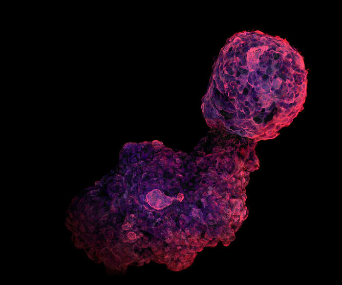
Image credits: © Syed Ashraf, Dr. Divya Sridharan and Ms. Salvia Zafar | Nikon Small World
#66 Image Of Distinction: Stephanie Huang, Victoria University Of Wellington, School Of Biological Sciences; School Of Psychology, Wellington, New Zealand
“Pyramidal neurons from the ventral orbital cortex (prefrontal cortex) from an adult rat brain.”
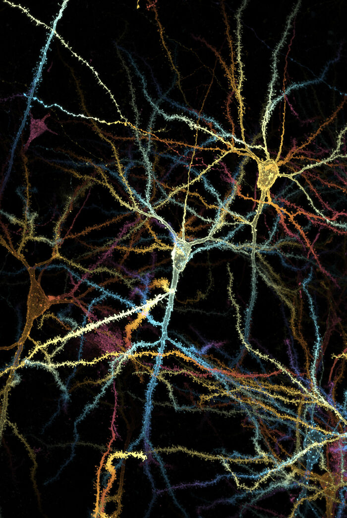
Image credits: © Stephanie Huang | Nikon Small World
#67 Image Of Distinction: Thomas Barlow, Sergio Bernal-Garcia And Kevin Gonzalez, Columbia University, Department Of Neurobiology And Behavior, New York, New York, USA
“Mouse pyramidal neuron, from the hippocampal CA1 region.”
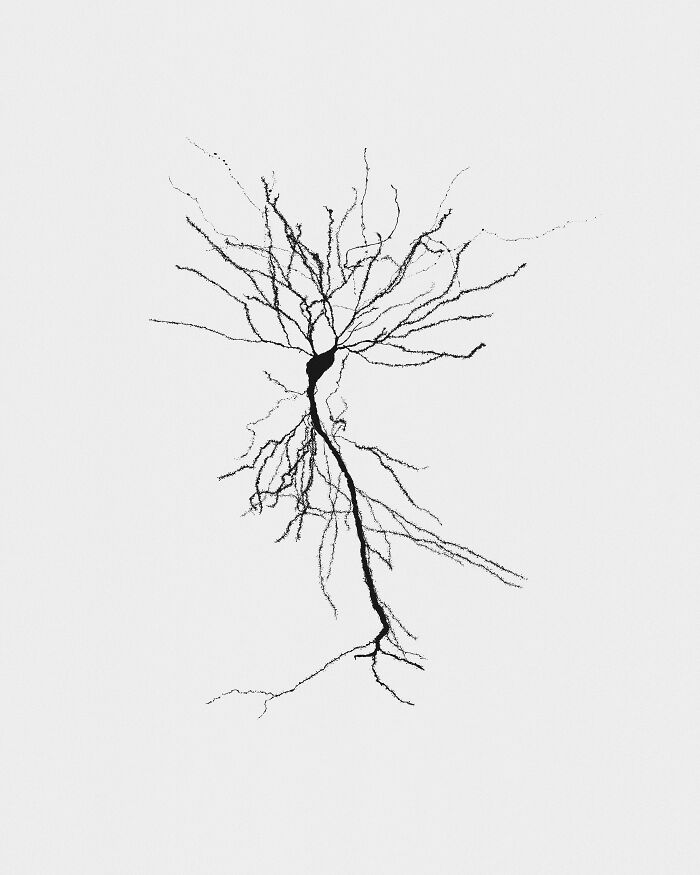
Image credits: © Thomas Barlow, Sergio Bernal-Garcia and Kevin Gonzalez | Nikon Small World
#68 7th Place: Stella Whittaker, National Institutes Of Health, National Institute Of Neurological Disorders And Stroke, Bethesda, Maryland, USA
“iPSC-derived sensory neurons labelled to show tubulin and actin.”
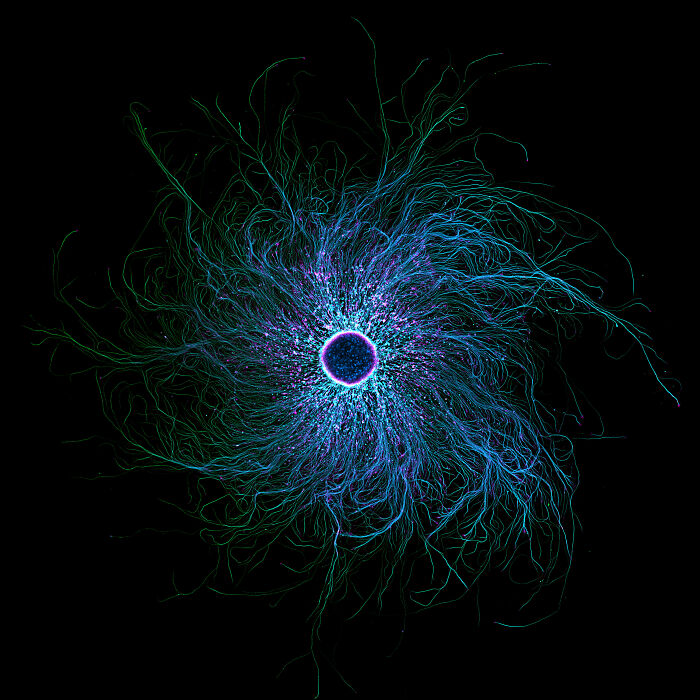
Image credits: © Stella Whittaker | Nikon Small World
#69 12th Place: Halli Lindamood And Eric Vitriol, Augusta University, Department Of Neuroscience And Regenerative Medicine, Augusta, Georgia, USA
“The actin cytoskeleton (cyan) and endoplasmic reticulum (red) of a mouse brain cancer cell.”
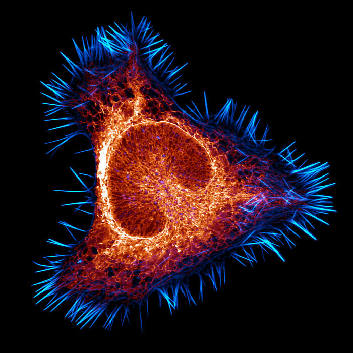
Image credits: © Halli Lindamood | Nikon Small World
#70 17th Place: Hong Guo, Chengdu, Si Chuan, China
“Water fleas (Daphnia) and algae.”
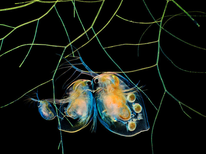
Image credits: © Hong Guo | Nikon Small World
#71 18th Place: Marius Mählen, Koen Oost, Prisca Liberali And Laurent Gelman, Friedrich Miescher Institute For Biomedical Research, Basel, Basel Stadt, Switzerland
“Fluorescently marked mouse colon.”
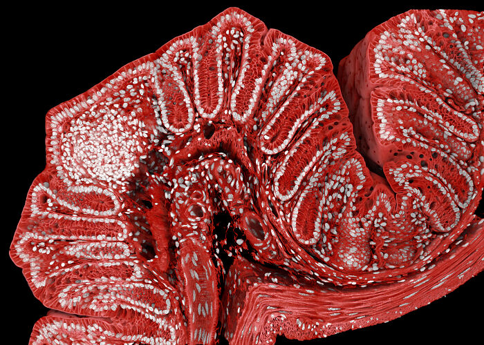
Image credits: © Marius Mählen, Koen Oost, Prisca Liberali and Laurent Gelman | Nikon Small World
from Bored Panda https://ift.tt/HPQIlnT
via IFTTT source site : boredpanda